


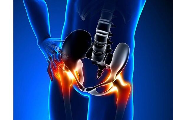
Osteoarthritis of the hip joint (Coxarthrosis)- It is a chronic degenerative joint disease, which leads to the deformation of the bone tissue.With coksartrose, all the components of the joint are involved in the pathological process: articular cartilage, bone structures adjacent to cartilage, synovial shell, ligaments, capsule and adjacent muscles.In the event of illness, the articular cartilage is destroyed, micro-sreeds of bones and osteophytes (bone growth) appear and inflammation of the ligamentous muscle of the hip joint occurs.
In the world, each fifth person complains of joint problems with the joints.It can be both pain or restriction of movement in the joints and a combination of these symptoms.Each second ambulatory vision lies with patients with bone muscle disorders, while 66% of cases are people under 65.According to the latest epidemiological research, the prevalence of osteoarthritis of the knee and hip joints among the adult population is 13%.
Risk factors for the development of coxarthrosis:
- Genetic predisposition.A common cause of coksartrose of the hip joints is the congenital or acquired mutation of the type II prollagen type.
- Age of the elderly.The probable cause of the prevalence of osteoarthritis in old age is a difference between the harmful effect on the articular cartilage of the external environment and its catering capacities.
- Ground.Women suffer more often from osteoarthritis than men.This is due to the effects of the influence of female sex hormones of estrogens on bone-mineral metabolism.However, the influence of the soil is ambiguous - according to some authors, unlike damage to other joints, there are no differences in the sexual base of coksartrose: in men, osteoarthritis of hip joint is as often found as in women.
- Excess body weight.The relationship is proven between excess body mass and the occurrence of osteoarthritis.The excess adhesive fabric increases the damaging load on the cartilage.In addition, adipose tissue produces pro-inflammatory enzymes that damage the cartilage fabric.
- Frequent development of bones and joints.In accordance with studies, 80% of coxarthrosis, which occurs for no apparent reason, is associated with previously not diagnosed defects in the development of hip joint - dysplasia and subluxation.
- Heavy physical work.Excessive load on hip joints with certain types of physical work can cause damage to the joints and the formation of osteoarthritis.Agricultural workers, diggers and people of similar work specialties.
- Injuries.The risk of developing coxarthrosis increases after a hip joint injury.In addition, an injured joint and both can be involved in the process.
- Professional sports.Professional sport can cause the occurrence of coxarthrosis at the same time due to the excessive burden on the joints and due to injuries.Potentially dangerous sports include heavy athletics, athletics jump, sport parachute.
- Joint bones and diseases- Rheumatoid arthritis, psoriatic arthritis, joint infections, avascular necrosis, goutuse arthritis, etc.
- Endocrine pathologies- Hypothyroidism, hypoparathyroidism, acromegaly (altered function of the anterior pituitary gland), diabetes, obesity.
If similar symptoms are detected, consult a doctor.Not self -medicate - it's dangerous for your health!
Hip joints' arthritis symptoms
The main symptoms of coxarthrosis include: pain, mobility restrictions and crunching it in the joints, their deformation, functional shortening of the lower limb and periodic swelling in the joints.
Pain of various intensities.The pain in the joint is initially insignificant and occurs for a short period.They appear or intensify by walking or with other physical efforts, for example, during squats, inclinations and weight.As the disease develops, the pain intensifies and even a long rest does not bring relief.In addition, the pain occurs with prolonged immobility and fixing the joint in a position.
Patients complain about “start-up” pain "SO-SOKED“ start-up ”pain in the hip joints after sleeping, leading to a car and other prolonged immobility.The pain "to start" for coxarthrosis does not last more than 30 minutes.Pain intensifies during hypothermia or in a stressful situation.They can be located in the buttock or groin area, on the front or side surface of the thigh.With the spread of pain on the nerves of the lumbar plexus, it can be transmitted to the thighs far from the center of the body or knee.Sometimes the pain applies to the Lombo-Sacred spine and the coccyx.
Restriction of joint mobility.The hip joint movements with Coksartrose are limited due to pain.At the same time, rotation (turns inside and outside) and bringing the lower limb (movement in the middle of the body) are more often disturbed, but can be limited (movement from the average axis of the body), as well as flexion and extension.The inability to make passive movements in the joint due to a pronounced pain syndrome causes a compensatory pelvic bias.The patient's approach changes, the buttocks retreat, the body deviates when transferring weights on the damaged side.With bilateral damage in patients with coksartrose, a "duck approach" is formed.
With coxarthrosis periodically occursSwelling in the jointwhich can be invisible due to the muscle layer and fat.In addition, the disease is characteristicCryst in the joints during movement, their progressive deformation and their functional shortening of the lower limb.
Often, a joint is affected by the disease, then the process applies to others.But sometimes osteoarthritis affects several joints at a time and polyosostoarthritis occurs.Polyosteoarthrosis is characteristic of the elderly or with a hereditary predisposition and concomitant diseases - bone diseases, joints and endocrine disorders.
Pathogenesis of osteoarthritis of the hip joints
In the pathogenesis of osteoarthritis of the hip joints, an important role is played by mechanical damage (injuries and microtraumas due to increased physical effort on the joint) and genetic, hormonal and metabolic factors.Often, it is not possible to discover which factor has influenced the development of the disease in a particular patient, but often the disease develops after tissue lesions with mechanical lesions.
Fabric damage stimulates the division of tissue cells from cartilage (chondrocytes), while the production of pro-inflammatory cytokines increases, which are normally present in cartilage in small quantities.Cytokines launch the inflammatory process, for example, under the influence of the IL-1 pro-inflammatory cytokine, enzymes are distinguished which destroy the cartilage of the joint.In addition, under the influence of cytokines, the production of the Enzyme Tsog-2 and other substances which have a toxic effect on the increases in cartilage.
Synovitis also play an important role in the development of coxarthrosis - inflammatory diseases of the synovial shell of joints or ligaments with the accumulation of liquid in the cavity.
A decrease in the elasticity and resistance of the articular cartilage associated with metabolic disorders leads to a decrease in its resistance to mechanical stress.With Coksartrose, all the components of the joints are involved in the pathological process, including an under-subcounted bone.Due to the fact that the large joints of the lower limbs represent the great joints of the body, they experience an important mechanical constraint, because of which microvalias occur in the subchondral plate and cartilage.Following microvèlates, the subchondral bone is compacted, which leads to the regional growth of osteophytes bone tissue.And this, in turn, stimulates an additional degradation of the articular cartilage.
In some cases, osteoarthritis of the hip joint is inherited.Hereditary osteoarthritis is supposed to be the polygenic heritage - due to the action of many genes, each of which affects weakly.The cause of certain diseases is a mutation in genes coding for macromolecules of joint cartilage, which causes its ruptures.The genes responsible for the chondrocytes division can also suffer from it.In addition, metabolic disorders are inherited, such as pyrophosphate arthropathy - a disease in which calcium pyrophosphate crystals accumulate in joint cartilage and synovial fluid.
Classification and stages of the development of osteoarthritis of the hip joints
According to the causes of the disease, coxarthrosis is divided into two main forms: primary (idiopathic) and secondary (resulting from or because of other diseases).
Primary coksartrosis:
- Located (only hip joints affect):
- unilateral;
- bilateral.
- Generalized (polyosteoarthrosis) with a lesion of at least three joint groups (for example, the hip, the knee and the small joints of brushes or feet).
Secondary osteoarthritis:
- Post -trauma:
- acute - due to an acute injury;
- Chronicle - Due to classes of certain sports or following a professional activity.
- Metabolic diseases (oconosis, hemochromatosis, Wilson disease, left -handed disease).
- Congenital pathologies and development defects (congenital dysplasia of the hip joint, relevant the disease, the sliding of the epiphysis of the femur, hypermobility syndrome, shortening of the lower limb, scoliosis, bone dysplasia).
- Endocrine pathologies (acromegaly, hypothyroidism, diabetes mellitus, hyperparathyroidism, obesity).
- Calcium bells (pyrophosphate arthropathy, calcifying tendonitis).
- Diseases of bones and joints (rheumatoid arthritis, psoriatic arthritis, soset disease, avascular necrosis, infections).
According to clinical manifestations, the following forms of coxarthrosis are distinguished:
- Few symptoms.
- Manifesto, manifested by brilliant clinical symptoms:
- Quickly progressive, in which symptoms develop in the first four years from the appearance of the disease;
- Slowly progressive - clinically significant symptoms appear after five years of the disease.
In accordance with the X-Ray image, two types of hip joints can be identified::
- Hypertrophic - with signs of increased restorative response (lesions are replaced by a new fabric, for example, osteophytes appear);
- Atrophic (decrease in tissue volume).
The stages of the disease can be determined radiologically and clinically.To determine the radiological stage of osteoarthritis of the hip joint, the classification of Kellgren and Lawrence (1957) is most often used.
Steps of osteoarthritis in radiological classification
| Scene | Signs |
|---|---|
| 0 | There is no sign of osteoarthritis in x -ray images |
| 1 | The joint space is not modified, the single regional osteophytes are visualized |
| 2 | The joint gap is not modified, significant regional osteophytes are visualized |
| 3 | The height of the joint space is moderately reduced, significant regional osteophytes are visualized |
| 4 | The height of the joint space is considerably reduced, significant regional osteophytes and subchondral osteosclerosis are visualized (compacting of bone tissues below the lower surface of the cartilage with the structure of the cartilage) |
To determine the clinical stage of the disease, classification (1961) is used, which uses both clinical signs and visualization criteria.
Osteoarthritis clinical stages
| Scene | Signs |
|---|---|
| 0 | The joint space is narrowed unequivocally and unevenly, the edges of the joint cracks are slightly pointed (initial osteophytes), a slight restriction of the movements is noted |
| 1 | The joint difference is considerably narrowed (50-60%), significant osteophytes, subchondral osteocosclerosis and cystic illumination in bone epiphysis;The clinic is predominant by the restriction of mobility in the joints, a rough crisis during movements, insignificant or moderate muscular atrophy |
| 2 | deformation, stiffness of the joint;The joint gap is narrowed by more than 60 to 70% of the standard or osteophytes extended completely absent, subchondral cysts, joint "mice" are visualized, cartilage or mixed pathological formations located in the joint cavity |
Complications of hip joints' arthritis
With coxarthrosis, all complications are precisely associated with pathological changes in the joints.
The course of coksartrosis can be complicated by local inflammatory processes:
- Stock Exchange - Inflammation of synovial bags in the joints;
- Tendovaginitis - Inflammation of the inner shell of the vagina of muscle tendons;
- Tunnel-printing syndrome of the nerve due to the formation of large osteophytes or joint deformation.
With the progression of coxarthrosis and its transition to clinical stages II and III, pain limits the mobility of the joint, and over time, joint ankylosis (fibrous, bone or cartilage) occurs, accompanied by its complete immobility.
Significant joint deformation can lead toFractures or aseptic necrosis of bones.For coksartrosis, the aseptic necrosis of the femoral head is the most formidable complication.
With pronounced coksartrosis, can occursubluxation and dislocation of the jointas well as the penetration of the femoral head into the pelvic cavity.The dislocations and the subluxation of the hip joint cause pain (first acute, then dull and painful), intensify during walking and other physical efforts, as well as the deformation of the joint, blade and sometimes to shorten the affected limb.
Despite the lack of systemic manifestations of osteoarthritis itself, in modern clinical practice, more attention is paid to the diseases associated with it.These are such pathological conditions that exist or arise in the context of current disease.In connection with inflammatory reactions on osteoarthritis, the formation of atherosclerotic plates on the internal walls of the vessels is improved, which increases the riskCardiovascular diseases.A decrease in physical activity due to pain and the restriction of joint mobility leads toObesity, depression and deterioration of quality of life.With prolonged use of non-steroidal anti-inflammatory drugs,The upper gastrointestinal cuts are affected,And alsoThe risk of cardiovascular pathologies and kidney diseases increases.
Diagnosis of osteoarthritis of the hip joints
The diagnosis of "coksartrose" is made on the basis of clinical manifestations and radiological examination.There is no characteristic laboratory sign for the diagnosis of osteoarthritis.
Among the clinical manifestationsThe main diagnosis of osteoarthritis of the hip joint is pain and its character.Pain for osteoarthritis of the hip joint occurs and is developing gradually over several years (sometimes several months with a quickly progressive form).The pain occurs or improves during physical effort or as a standing position.If the patient begins to feel pain alone, then inflammation (synovitis) has joined.The declaration is noted until 30 minutes in the morning and with prolonged immobility.
The limitation of joint mobility increases gradually, which applies to active and passive movements.With the development of the disease, the joints are deformed, the functional shortening of the length of the limbs can occur.
To a physicine examinationThere is a limitation of joint mobility, their deformation, shortening of the limbs, pain on the palpation of the joint and a large spinning of the femur, a muscular atrophy.
Laboratory methodsFor the diagnosis of osteoarthritis of the hip joints, it is not necessary.However, they can be used for the differential diagnosis of coxarthrosis with arthritis (rheumatoid and chronic), because with osteoarthritis, there are no inflammatory changes in the overall blood test and the rheumatoid factor, and the levels of uric acid are not increased.In addition, using laboratory tests, contraindications are revealed for drug treatment methods.
Instrumental methodsFor the diagnosis of osteoarthritis of the hip joints:
- Radiography- This is the main method of diagnosing osteoarthritis of the hip joints.The radiography determines the characteristic changes of coksartrosis: narrowing of the joint, osteophyte, erosion and ulceration of cartilage, subchondral cysts and osteosclerosis.The X-Ray examination is a classic method for the diagnosis of coxarthrosis, and the radiological signs underlie the classification of coxarthrosis.However, at present, other methods of visualization of the articulation are increasingly used, such as imaging by ultrasound and magnetic resonance.
- Ultrasound examination (ultrasound) -The advantage of ultrasound is in the absence of a radial load on the body.
- Magnetic resonance tomography (MRI)- In comparison with other methods, it allows you to visualize joint lesions more clearly.
- Arthroscopy-Allows you to identify articular cartilage damage: chondrotation zones (softening of the articular cartilage) with a diameter of less than 10 mm to deep cracks which penetrate to the subchondral bone and the formation of deep ulcers.Superficial and medium cracks and surface erosion can also be viewed.
The identification of coksartrosis does not generally represent particular difficulties, but when evaluating a specific clinical situation, it is necessary to remember the possible secondary origin of the hip joints (as complications of other diseases, for example, with endocrine disorders).
Treatment of hip joints osteoarthritis
The treatment of osteoarthritis of the hip joints can be both conservative (drug and non -university) or operational.Conservative treatment is used at 1-2 stages of the disease, surgical-at 3 stages.Surgical treatment can be recommended at 2 steps with persistent pain and a lack of reaction to conservative therapy.
The objectives of conservative therapy:
- Improve quality of life - Reduce pain and increase joint mobility;
- Stop or slow down the development of the disease.
Non -drug treatment methods include:
- unloading of the hip joint (decrease in body weight, creation of additional support and transfer of part of the body weight to cane or crutches);
- Physiotherapy physiotherapy;
- Physiotherapeutic treatment methods.
The treatment of coxarthrosis begins with non -drug methods, an important role is given to physiotherapy exercises.With intense pain, the patient must use support.With a pronounced disease and the presence of endoprothetic contraindications, support must be used for life.
Cuxartrose medicinal therapyIncludes drugs that reduce symptoms of the disease.These are pain relievers, as well as drugs from the group of non-steroidal anti-inflammatory drugs (NSAIDs).NSAIDs are divided in non-election and selective.
Analgesics and NSAIDs for osteoarthritis of the hip joint are used for a short period to relieve pain and inflammation.Currently, there is no proven advantage of a non-steroidal anti-inflammatory agent compared to another, so that the choice of a particular drug depends on the side effects and a specific clinical situation caused by it.
It should be remembered that NSAIDs have a number of side effects.When you take them, the mucosa of the stomach and duodenum is affected, as a result of which ulcers and bleeding is possible.A number of NSAIDs have a toxic effect on the liver and kidneys.In addition, NSAIDs disrupt platelet aggregation and, therefore, the patient is disturbed by thrombosis and there is a tendency to bleed.NSAIDs for prolonged use suppress hematopoiesis processes and can cause aplastic anemia and agranulocytosis.The reception of selective NSAIDs causes complications much less.
The ointments and gels used locally cause fewer side effects than oral products.For the treatment of osteoarthritis, drugs with warming and pain reduction are used.They can contain turpentine, menthol, nicotinic acid esters, salicylates, bee venom.In addition, NSAIDs have a good effect.
In the absence of the effect of pain relievers and NSAIDs or if it is impossible to choose the optimal dose of the drug, the analgesics of central action can be prescribed in the short term.
In case of inflammation, intra-articular administration of corticosteroids is used.Corticosteroids are not used more than 2 to 3 times a year, as more frequent use can lead to degeneration of cartilage.
Drugs acting slowly weaken the symptoms of the disease include chondroprotectors, inappropriate compounds of lawyers or soy, hyaluronic acid.These drugs are included in the recommendations of the European anti -function league for the treatment of osteoarthritis of the hip joints.Preparations reduce pain and improve joint mobility.
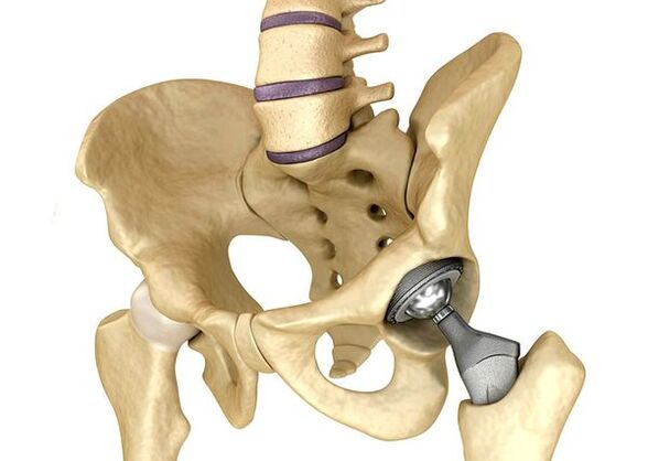
Hip joint endoprostheticIt is used in serious cases of stage III, when pain syndrome cannot be eliminated and the mobility of the joint is considerably limited.The hip joint prostheses lead to a decrease in pain syndrome, an improvement in the functional condition of the joint and the quality of the patient's life.The effect persists for 10 to 15 years, after which a second operation may be necessary.During surgery, hip joint is replaced by the artificial imitation of ceramics, metal (most often titanium prostheses) or polymer.
Forecast.Prevention
The prognosis for osteoarthritis of the hip joints in relation to the patient's life is favorable, but the disease often leads to a handicap.According to the World Health Organization, 80% of elderly patients with coxarthrosis have a violation of mobility and 25% cannot do daily things.In this regard, the primary prevention of osteoarthritis of the hip joints is important.
Prevention measures:

- Reduce body weight.It is necessary to adjust nutrition to reduce weight and load on the joint.In addition, a decrease in the volume of adipose tissue reduces the quantity of inflammation mediators that he has released.
- Avoid heavy and sports physical overloads.Physical overloads are often the cause of osteoarthritis of the hip joints, while moderate physical activity, on the contrary, improves the condition of articular cartilage, retains its normal mobility and reduces load on other joints.
- Correct the underlying disease.If the patient is detected in diseases that can lead to secondary coksartrosis (endocrine, rheumatic and others), the underlying disease is necessary.The normalization of the hormonal context and the realization of a persistent remission of rheumatic diseases are both the main prevention of osteoarthritis and allows you to slow down its development.
- Lead a healthy lifestyle.A balanced diet with a sufficient content of vegetable and animal protein, polyunsaturated fatty acids and limiting simple carbohydrates, as well as moderate physical activity, avoid the occurrence of coxarthrosis even in the presence of risk factors.
Currently, prevention of hip joint disease is compulsory in neonatology and pediatrics.Over time, congenital dysplasia adjusted from the hip joint considerably reduces the risk of coxarthrosis in adulthood.